For centuries, people have been fascinated with the human body and measuring its dimensions, believing that an individual’s physical health and performance is closely linked to their body size and shape. In the early days, physicians such as Hippocrates (460-370 B.C.) used subjective descriptions of a person’s physical build to predict their risk of developing certain diseases, such as tuberculosis and stroke. Thankfully, there have been extraordinary advances in the tools used within medical practice since then, along with the creation of more quantitative methods for measuring the human body, otherwise known as anthropometry. However, though the field of anthropometry has seen major developments since its earliest origins, the tools which are regularly used to measure the human body, in fields such as fashion and sport, are the same as those that have been used for the past century. These traditional techniques rely on the use of tape measures, height gauges and weight scales, with combinations of these used to create simple proxies of weight status and body shape, such as the body-mass-index (BMI). These approaches for assessing the human body are prone to error and limited by their relative simplicity, as they do not capture its complex three-dimensional (3D) variations in shape and mass distribution.
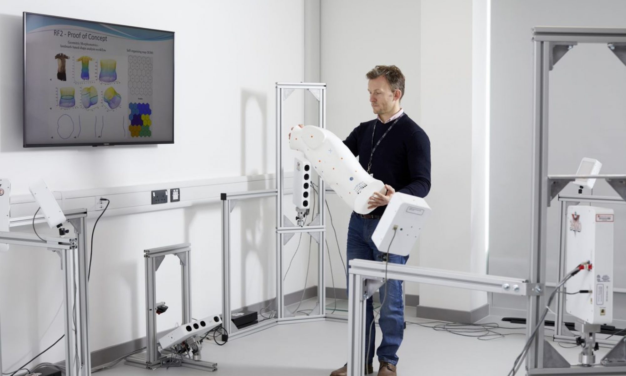
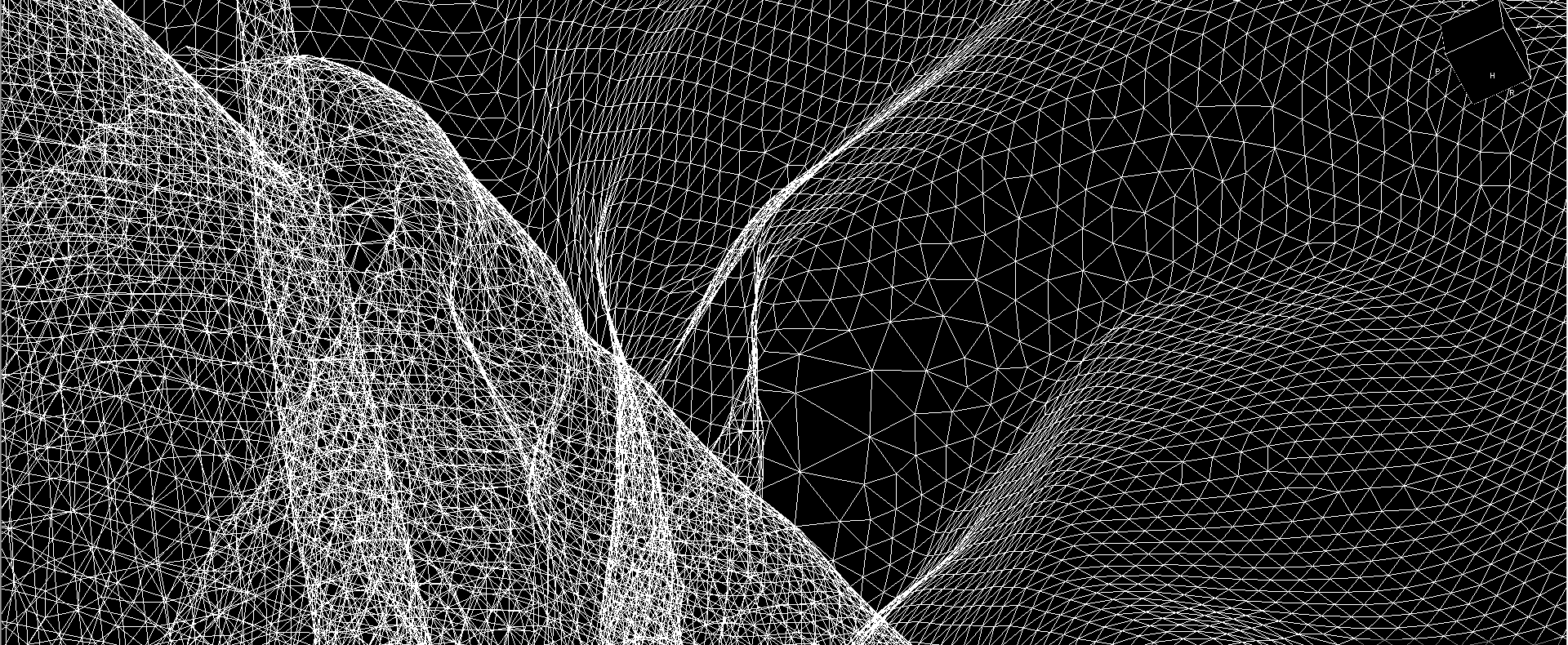
Since 2011, Sheffield Hallam University’s Morphology Research Group has become an international leader in the development of novel 3D imaging techniques for measuring and analysing the human body within elite sport, with morphology now one of the key research themes within the Sports Engineering Research Group (SERG). Modern 3D imaging systems, acquire point cloud data that captures detailed and accurate external dimensions and shape characteristics of the human body. An example of the Morphology Group’s work using 3D imaging in sport has been obtaining more accurate person-specific body segment inertial parameters (BSIPs) – e.g., mass, centre of mass position, moments of inertia. These metrics are vital when performing biomechanical analyses used to model human movement, particularly in movements involving high accelerations and rotations in acrobatic sports, such as trampolining and gymnastics. After scanning individuals using a 3D imaging system, Choppin et al., (2020) were able to calculate the magnitude and orientation of principal moments of volume from the resulting geometries, demonstrating the potential for errors when calculating these variables using more traditional techniques. It was also suggested that low-cost 3D imaging devices could offer a viable alternative solution for obtaining BSIP’s when possible.

Size Stream LLC booth scanner.
More recently, the Morphology Research Group has been working to translate this knowledge, which has been developed within sport regarding new measures of body shape derived from 3D imaging data, into health and wellbeing applications, such as chemotherapy dosing. Currently, estimates of body surface area (BSA) are used to determine a person’s drug dosage when receiving conventional chemotherapy as part of their cancer treatment. Though this method has been used extensively during the past century, the continued use of BSA-based dosing remains controversial due to these methods of calculation having limited validity, particularly with modern populations that tend to have higher BMI values. The implications of these inaccuracies in dose calculation are significant since it may reduce both the effectiveness of a person’s treatment and their ability to tolerate it safely. Consequently, calls for improved methods of dose calculation have increased in recent years and remains a significant challenge within oncology.
The Morphology Research Group recently published a new paper which presents the idea that the use of advanced body measurement techniques, such as 3D imaging, can provide oncology practitioners with improved tools for prescribing chemotherapy dosages that are valid for individuals, regardless of their body type. Though 3D imaging devices have previously been too large and expensive to be feasibly used in practice, recent developments in low-cost, smartphone-based systems that can acquire 3D imaging data have led to increased accessibility of this technology. Studies to develop improved methods for determining chemotherapy dosages for people with atypical body types using these advanced body measurement techniques are currently ongoing.

me°- three – sixty scanning app from Size Stream LLC
This blog article is taken from the SERG blog.
To find out more information about SERG and the Morphology Research Group’s work, check out our website, our annual review or our MSc Sports Engineering course. You can also follow us through our social media channels, available at the top right of this page or through our linktr.
References:
Thelwell, M.; Masters, N.; Appleyard, R.; Bullas, A.M. Advanced Body Measurement Techniques Can Complement Current Methods of Cytotoxic Chemotherapy Dose Prescription. Appl. Sci. 2024, 14, 834. https://doi.org/10.3390/app14020834
Choppin, S., Clarkson, S., Bullas, A., Thelwell, M., Heller, B., Wheat, J. Anatomical and principal axes are not aligned in the torso: Considerations for users of geometric modelling methods. Journal of Biomechanics, Volume 114, 2021, 110151, https://doi.org/10.1016/j.jbiomech.2020.110151.